
|
News Detail
Orthopedic Beauty: Anatomy of the Subtalar Joint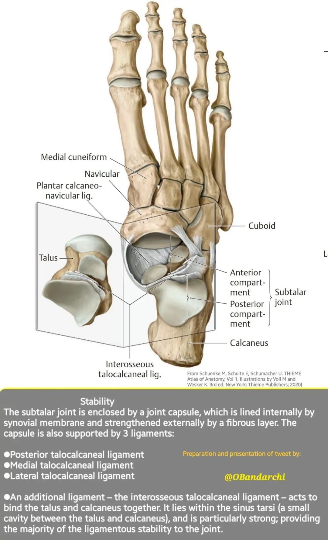
The subtalar joint is a joint between two of the tarsal bones in the foot – the talus and the calcaneus. Tarsal bones are the seven bones that form the heel, ankle, and back of the foot. The talus is the bone that connects the ankle to the foot, and the calcaneus is the heel bone. The subtalar joint is also known as the talocalcaneal joint. The subtalar joint is classified structurally as a synovial joint, which means that it has a fluid-filled capsule that surrounds the bones and allows smooth movement. The joint is also classified functionally as a plane synovial joint, which means that it allows gliding movements in different directions. The subtalar joint is the main joint that allows inversion and eversion of the foot. Inversion is the movement of turning the sole of the foot inward, and eversion is the movement of turning the sole of the foot outward. These movements are important for adapting to uneven surfaces and changing directions. The subtalar joint also contributes to other movements of the foot, such as plantarflexion (pointing the toes down) and dorsiflexion (lifting the toes up), but these movements are mainly controlled by other joints in the foot and ankle. The subtalar joint has three articular surfaces, or flat areas where the bones meet. These are:
The subtalar joint is held together by several ligaments, which are strong bands of connective tissue that connect bones. The main ligament that stabilizes the subtalar joint is the interosseous talocalcaneal ligament, which runs along a groove between the talus and calcaneus. Other ligaments that support the subtalar joint are:
The subtalar joint is innervated by nerves that carry sensory and motor information to and from the brain. The nerves that supply the subtalar joint are:
The subtalar joint is vascularized by blood vessels that deliver oxygen and nutrients to the tissues and remove waste products. The blood vessels that supply the subtalar joint are:
The subtalar joint can be affected by various injuries and conditions, such as sprains, fractures, arthritis, infections, tumors, or congenital deformities. Some common symptoms of subtalar joint problems are pain, swelling, stiffness, reduced range of motion, instability, or deformity of the foot. Treatment options depend on the cause and severity of the problem, but may include rest, ice, compression, elevation, medication, physical therapy, braces, orthotics, injections, or surgery.
|

Shanghai Carefix Medical Instrument Co., Ltd.China
滬ICP備16042301號(hào)-1 (滬)-非經(jīng)營(yíng)性-2016-0122 滬食藥監(jiān)械生產(chǎn)許20101725號(hào)
