療器械有限公司")
|
News Detail
Ring fixator (Ilizarov)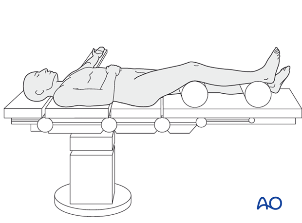 1. Principles and indicationsPrinciplesAs the ring fixator is an external fixator, it gives relative stability. IndicationsIn fresh fractures, there are several indications for using a ring fixator:
2. Patient preparationThis procedure is normally performed with the patient in a supine position for ring fixator. 3. Safe zones for pin insertionInserting percutaneous instrumentation through safe zones reduces the risk of damage to neurovascular structures.  4. FixationProximal ring placementThe proximal ring is placed at the level of the head of the fibula and parallel to the knee joint.  Distal ring placementPlace the ring at the level of the proximal end of the syndesmosis.  First intermediate ringAdd a second ring in the proximal fragment of the midshaft, connecting it with 4 rods to the proximal ring. Note:  Second intermediate ringAdd a second ring in the distal fragment of the midshaft, connecting it with 4 rods to the distal ring.  5. Reduction and final fixationConnect the two intermediate rings with 4 rods without completely tightening the bolts.  6. Aftercare following application of a ring fixatorImmediate postoperative careImmediately after surgery, while the patient is still in the hospital, emphasis is given to:
The patient’s leg should be slightly elevated, with the leg placed on a pillow, 4 cm above the level of the heart. Advise the patient about foot positioning in order to avoid equinus deformity.  Pin-site careProper pin/wire insertion
Pin-site care
Pin/wire loosening or pin tract infection
Before changing to a definitive internal fixation an infected pin tract needs to heal. Otherwise infection will result. MobilizationImmediately postoperatively, all joints (hip, knee, ankle) are actively mobilized.  Weight bearingPartial weight-bearing with crutches should begin as soon as possible. Depending on the consolidation, weight bearing can be increased after 6-8 weeks with full weight bearing when the fracture has healed.  Follow upClinical and radiological follow-up is recommended after 2, 6 and 12 weeks. Implant removalRemove the fixator after clinical and radiographical bony healing.
|

Shanghai Carefix Medical Instrument Co., Ltd.China
滬ICP備16042301號-1 (滬)-非經(jīng)營性-2016-0122 滬食藥監(jiān)械生產(chǎn)許20101725號
