Shanghai CareFix Medical Instrument Co., Ltd.
News Detail

Intraoperative Fluoroscopic Techniques for Tibial Plateau Fractures
文章附圖
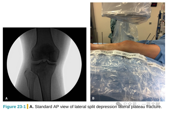
  1. Standard Anteroposterior (AP) View: The patella is located between the medial and lateral femoral condyles, with 1/3 to 1/2 of the fibular head overlapping the lateral margin of the tibial plateau.
    1.1 External Rotation View: The patella is positioned over the lateral femoral condyle, with increased overlap between the proximal tibia and fibula.
    1.2 Internal Rotation View: The patella is positioned over the medial femoral condyle, with decreased or no overlap between the proximal tibia and fibula.
  2. Head-Tilted 10° AP View: On the basis of the standard AP view, tilting the C-arm fluoroscope head by 10° can neutralize the posterior slope of the tibial plateau articular surface. As a result, the X-ray beam is parallel to the articular surface, which appears linear. For comparison, refer to the following figures: Figure 23-1 shows the standard AP view, while Figure 23-2 shows the head-tilted 10° AP view. Note the differences in the presentation of the tibial plateau articular surface.
  3. Standard Lateral View: The medial and lateral femoral condyles are completely superimposed, with the lateral tibial plateau articular surface higher than the medial plateau.
    As shown in the following figure: In the lateral view after a split and collapse of the lateral plateau, the collapsed lateral articular surface (indicated by the yellow arrow) is lower than the intact medial plateau (indicated by the blue arrow).

  4. Modified Lateral Views:
    4.1 Varus Lateral View: Elevating the medial plateau to assess the reduction of the medial plateau articular surface.
    As shown in the following figure:
    4.2 Valgus Lateral View: Elevating the lateral plateau to assess the reduction of the lateral plateau articular surface.
    As shown in the following figure:


Login
Login
Login by:
leave a message
back to the top
91在线资源_先锋在线资源_少妇特黄A一区二区三区_国产一级二级三级