
|
News Detail
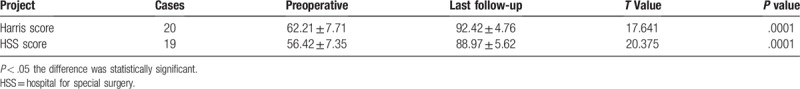
Successful management of lower limb nonunion using locking plates and bone graft with retention of i As a library, NLM provides access to scientific literature. Inclusion in an NLM database does not imply endorsement of, or agreement with, the contents by NLM or the National Institutes of Health. Abstract This study aims to investigate the clinical effect of the combined an additional locking plate with bone graft based on retaining the original intramedullary nail for the treatment of lower limb nonunion. From June 2008 to December 2012, 39 patients were admitted and treated, who developed non-infectious bone nonunion after intramedullary nail fixation for long bone fracture in the lower limb. Additional locking plate and autogenous iliac bone grafting were performed for these patients, in which the original intramedullary nail was retained. Follow-ups were performed once at postoperative months 1, 2, 3, 6, and 12, and every year onwards. During these follow-ups, imaging and clinical function examinations were performed, in order to observe callus growth and the fractured limb functions. All patients have been followed-up, in which the duration of these follow-ups ranged between 8 and 24 months. All patients gained bony union within 6 to 11 months, and the healing rate was 100%. Radiographic healing time ranged between 8 and 15 months. Full weight-bearing time ranged between 2 and 10 months. According to Harris hip scores and Hospital for Special Surgery (HSS) Knee joint scores, 17 cases were excellent, 2 cases were good, and 1 case was acceptable; with an excellent and good rate of 95.00%. According to HHS score for the knee, 15 cases were excellent, 3 cases were good, and 1 case was acceptable; with an excellent and good rate of 94.74%. The combined treatment of the additional blocking plate with bone grafting based on retaining the original intramedullary nail for bone nonunion could effectively eliminate lateral and rotatory instability of the fractured ends. This surgical method had a short operation time, high healing rate and other advantages. Keywords: 1.?Introduction Due to advantages such as central fixation, small trauma and high union rate, interlocking intramedullary nail fixation treatment for long bone diaphyseal fractures has been widely used in clinical practice. However, after intramedullary nailing, patients would develop varying degrees of lateral instability; and the incidence of nonunion is approximately 1.8%.[1] 2.?Materials and methods 2.1. General information From June 2008 to December 2012, 39 patients who developed non-infectious bone nonunion after intramedullary nail fixation for long bone fractures in the lower extremity were admitted and treated. The study was approved by Medical Research Ethics Committee of Luoyang Orthopedic Hospital of Henan Province (approval number: 20080613). Among these patients, 28 patients were male and 11 patients were female. Fracture causes: 13 cases were due to traffic accidents, 11 cases were due to crushing, and 15 cases were due to falling from a high position. Fracture sites: fractures in 27 cases were located in the femur, wherein fractures in 6 cases were located in the upper tertile, fractures in 14 cases were located in the medium tertile, and fractures in 7 cases were located in the inferior tertile; fractures in 12 cases were located in the tibia, wherein fractures in 3 cases were located in the upper tertile, fractures in 6 cases were located in the medium tertile, and fractures in 3 cases were located in the inferior tertile; fractures in 33 cases were closed fractures, which were treated with reamed antegrade interlocking intramedullary nailing. Furthermore, 6 cases had open fractures (4 cases were Gustilo I, and 2 cases were Gustilo II), which were treated with emergency debridement followed by unreamed antegrade interlocking intramedullary nailing. Nonunions were classified by the Weber-Cech classification, according to the vitality of fractured fragments: 29 cases were hypertrophic type, and 10 cases were atrophic type. AO classification: 8 cases were type A2, 6 cases were type A3, 5 cases were type B1, 5 cases were type B2, 2 cases were type B3, 9 cases were type C1, and 4 cases were type C2. The intervals from fracture to nonunion ranged between 13 and 28 months, with an average interval of 19.6?±?4.3 months. 2.2. Diagnostic basis 1. At more than 9 months after intramedullary nailing, there was no evidence of any increase in bone callus in the last 3 months. 2. The presence of local tenderness in the fracture sites was found during the physical examination, and pain increased during weight-bearing or activities. 3. X-ray films exhibited hardening in the fracture ends, no continuous callus connection, or at least 3 lateral cortices on the lateral sides had no continuous callus, no main nail fracture, or internal fixation failure. 2.3. Selection criteria of disease cases Inclusion criteria: 1. patients who were between 18 and 60 years old; 2. patients who met the diagnostic criteria of nonunion after intramedullary nail fixation; 3. patients who agreed to retain the original intramedullary nail and undergo additional plate fixation, and provided a signed informed consent. Exclusion criteria: 1. patients with original fractures that were pathologic fractures; 2. patients with local soft tissue infection or infected nonunion; 3. patients who had serious heart, liver, lung, kidney diseases and other systemic diseases, and were not tolerant to surgery. 2.4. Surgical methods Combined spinal epidural anesthesia (CSEA) was performed. Patients were laid in the supine position. The skin, subcutaneous tissue and deep fascia were cut layer by layer at the original incision, the adhesion tissue was isolated, and the fractured ends of the femur or tibia were exposed. Different degrees of rotation or lateral abnormal activity were found during the operation. Fractured ends were cleaned, hyperplasia and free sequestrum in the fractured ends were removed, and the hardened bone was thoroughly scrapped with a curette. The callus and connective tissue at the sides of the surgical approach were removed to facilitate the placement of an additional steel plate, while too much stripping was not necessary in other directions. For the screws and steel plates, those made with the same material should be selected as much as possible. With the fractured end as the center, the steel plate was placed aside the diaphysis, biasing forwards or backwards. The drilling direction should be mastered while the screw was driven into the bone. The locking hole could be fixed with unicortical or bicortical locking nails in the axial position. The pressurized hole could be fixed with bicortical screws. The screw could slant forwards or backwards within a certain range, and stuffed through the contralateral cortical bone by the side of the intramedullary nail to avoid blockage. When the placement of the plate observed by X-ray was satisfactory, the incision was repeatedly and thoroughly rinsed, and was temporarily tied and covered with sterile dressing materials. According to bone defect size, an appropriate ipsilateral or contralateral ilium was obtained and prepared into matchstick-like strips. Before grafting, both sides of the cortical bone in the fractured end were slightly cut and stripped, in order to prepare a fresh fracture section. Then, the bone strips were closely embedded into the gap between the fracture ends and the 2 to 3?cm surrounding region, with a broad range; in order to increase the bone contact area. Bone strips were slightly compressed to prevent movement. When the bone grafting surgery was satisfactory, operative materials were counted, a negative pressure drainage tube was placed in the incision, and the incision was sutured layer by layer, and tied and fixed with sterile dressing. 2.5. Postoperative care and follow-up method After surgery, no patients underwent external fixation. Patients were guided to undergo functional training of the lower limb muscle groups on the first day after surgery. On the second day, the drainage tube was removed and CPM functional exercise was performed to prevent knee joint adhesions. When the X-ray image showed significant callus formation, full weight-bearing exercise was performed. 2.6. Evaluation criteria Fracture healing criteria: no local pain upon complete loading, no tenderness at the fracture, imaging reveals at least 3 cortical continuous calluses on the transverse and lateral sides of fractures, and no main nail fracture and fixation failure. Lower extremity hip scores based on Harris hip scores: excellent 90 to 100 points; good, 80 to 90 points; acceptable, 70 to 80 points; and poor, <70 points. Lower extremity knee joint scores based on HHS knee scores: excellent, more than 85 points; good, 70 to 84 points; acceptable, 60 to 69 points; and poor, <59 points. 2.7. Statistics analysis Data were analyzed using statistical software SPSS 16.0. Data were expressed as mean ± standard deviation, and preoperative and postoperative indicators were compared using paired 3.?Results 3.1. Surgical results In this study, operation time ranged between 60 and 110 minutes, with an average of 70.3?±?18.1?minutes. Intraoperative blood loss ranged between 120 and 230 ml, with an average of 135.2?±?39.4?ml. The duration of follow-up ranged between 8 and 24 months, with a median of 16 months. All incisions healed by first intention. After surgery, 22 patients had pains at the bone donor site, among which 20 patients achieved remission within 1 month, and 2 patients achieved remission after 3 months. All patients gained bony union within 6 to 11 months (average time was 8.1 months), and the rate of bony union was 100%. Radiographic healing time ranged between 8 and 15 months, with an average of 10.3 months. Full weight-bearing time ranged between 2 and 10 months, with an average of 5.2 months. Other interventions were not performed during the follow-ups, No plate fracture, infection, intramedullary nail loosing, or other complications occurred. 3.2. Evaluation of curative effects Average Harris hip joint scores in the 20 patients with femoral bone nonunion increased from 62.21?±?7.71 points before surgery to 92.42?±?4.76 points after surgery. In classifying according to HHS, 17 cases were excellent (85.00%), 2 cases were good (10.00%), and 1 case was acceptable (5.00%); with an excellent and good rate of 95.00%. The average HHS knee scores in seven patients with distal femur nonunion and 12 patients with tibia nonunion increased from 65.42?±?7.35 points before surgery to 88.97?±?5.62 points after surgery. In classifying according to HHS for knee scores, 15 cases were excellent (78.95%), 3 cases were good (15.79%), and 1 case was acceptable (5.26%); with an excellent and good rate of 94.74%. (Details are presented in Tables Table 1 Function score about Harris of lower limb hip joint and HSS of Knee joint
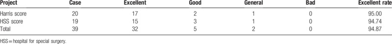
Table 2 Function evaluation about Harris of lower limb hip joint and HSS of Knee joint (case).
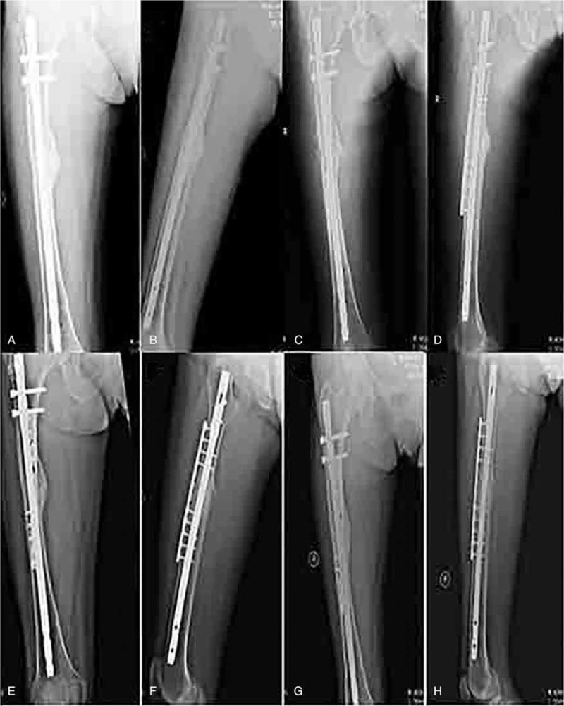
3.3. Typical case Patient A was a 43-year-old male patient. Bone nonunion occurred at the right middle femur after intramedullary nail fixation. The combined treatment with an additional steel plate and bone graft was performed on this patient. Continuous callus was found during re-examination at postoperative month 6, with blurring turning curves. The fracture was confirmed to heal during the re-examination at postoperative month 12 (Fig. HYPERLINK "https://www.ncbi.nlm.nih.gov/pmc/articles/PMC6455660/figure/F1/" \t "https://www.ncbi.nlm.nih.gov/pmc/articles/PMC6455660/figure"
Comparison of right femoral fractures before and after surgery. A: Preoperative positive position. B. Preoperative position. C. Positive position after operation. D. Posterior lateral position. E. Positive bitmap for 6 months after operation. F. Side map of 6 months after operation. G. Positive bitmap for 12 months after operation. H. Side map of 12 months after operation. 4.?Discussion Bone nonunion patients mostly have undergone many surgical treatments. Repeated surgical trauma results in great disease suffering and social psychological pressure. Hence, an operation method with less hospitalization expense, minimized trauma, and a high rate of fracture healing is particularly important. Rotation and lateral instability are the main reasons of nonunion after intramedullary nailing was performed in the lower limb fractures. At present, the commonly used surgical methods include: dynamization of the intramedullary nail; reamed intramedullary nailing; removal of the intramedullary nail plus plate fixation; plate fixation based on retaining the intramedullary nail. Dynamization of the intramedullary nail and reamed intramedullary nailing cannot solve the rotatory instability of fractured ends after intramedullary nail fixation, and dynamization may even further lead to local rotary instability, and may result to shortening of the affected limb. Due to its irregularity, the long bone marrow cavity cannot be effectively reamed by milling, and the instability problem of the fractured ends cannot be effectively solved by reaming the medullary cavity, resulting in a healing rate ranged from 78.3% to 100%.[9–13] The combined treatment with locking plate and autogenous iliac graft based on retaining the original intramedullary nail for low limb nonunion ensures axial stability, and eliminates local lateral and rotatory instability. The screws of the locking plate and steel plate form a complete system of mechanical support, which causes a longitudinal stress to travel through the neck of the nail directly to the fractured end along the plate, increases stability, provides a stable mechanical environment for the calcification healing of the fibrous cartilage, and improve fracture healing rate.[14–17] The difficulty of this operation is that the long bone marrow has to be fixed with the intramedullary nail. The pressurized plate would be locked using an additional steel plate with 6 to 11 holes, placed on the lateral side of the diaphysis, or slightly forwards. The drilling direction should be mastered when the screw is driven into the bone. The locking hole could be fixed with unicortical or bicortical locking nail in the axial position. The pressurized hole could be fixed with a bicortical screw to avoid blocking of the intramedullary nail. The screw could slant forwards or backwards within a certain range. During the operation, attention should be given to the drill tip to prevent screw breakage; and try to make use of the osseous pin, which is not easy to crack, in drilling holes. Bone grafting is extremely important in the treatment of bone nonunion; hence, bone grafting skill is also critical. 1. Preparation before the grafting: Before the bone grafting, fibrous tissues in the fractured ends should be thoroughly cleaned, and hardened cortical bones on both sides are cut and stripped, in order to establish fresh bone sections that could stimulate the local biological activities of the bone nonunion area, increase bone grafting area, and promote bone graft survival. 2. Source of the bone graft: Autogenous bones are preferred choices for grafts, and the autogenous iliac bone is considered to be the best source. The direct implantation of large bone blocks is not a good choice. The iliac bone should be prepared into matchstick-like strips, which are easy to implant and can better stimulate the osteogenesis chain effect. 3. The amount of bone grafts: The amount of bone graft should be sufficient. These would be absorbed if less bone is implanted, resulting in delayed union or ununion. 4. The range of the bone graft: The bone graft should not be confined within bone defect regions. The grafting range should be wide, and bone grafting should be conducted in both the proximal and distal fractured ends, in order to improve local bone quality and increase the osseous contact area. Autogenous cancellous bone contains abundant marrow stromal stem cells, which can induce and promote the formation of new bone, and promote the osteogenesis chain reaction. The majority of people believe that surgical indications of bone grafting for nonunion are the follows: bone defect is larger than 2?cm, or the defect area is larger than 50% of the diameter of the femur.[20] The combined treatment with additional locking plate and bone graft based on retaining the original intramedullary nail has many advantages, such as simple operation, minimal trauma, low cost of hospitalization, high fracture healing rate, and enabling early exercise. This treatment is an effective therapeutic method. Author contributions Conceptualization: Data curation: Formal analysis: Methodology: Resources: Validation: Writing – original draft: Writing – review & editing: Footnotes Abbreviations: CSEA = combined spinal epidural anesthesia, HSS = hospital for special surgery. The authors have no conflicts of interest to disclose. References [1] [2] [3] [4] [5] [6] [7] [8] [9] [10] [11] [12] [13] [14] [15] [16] [17] [18] [19] [20] [21]
|

Shanghai Carefix Medical Instrument Co., Ltd.China
滬ICP備16042301號-1 (滬)-非經(jīng)營性-2016-0122 滬食藥監(jiān)械生產(chǎn)許20101725號
