療器械有限公司")
|
News Detail
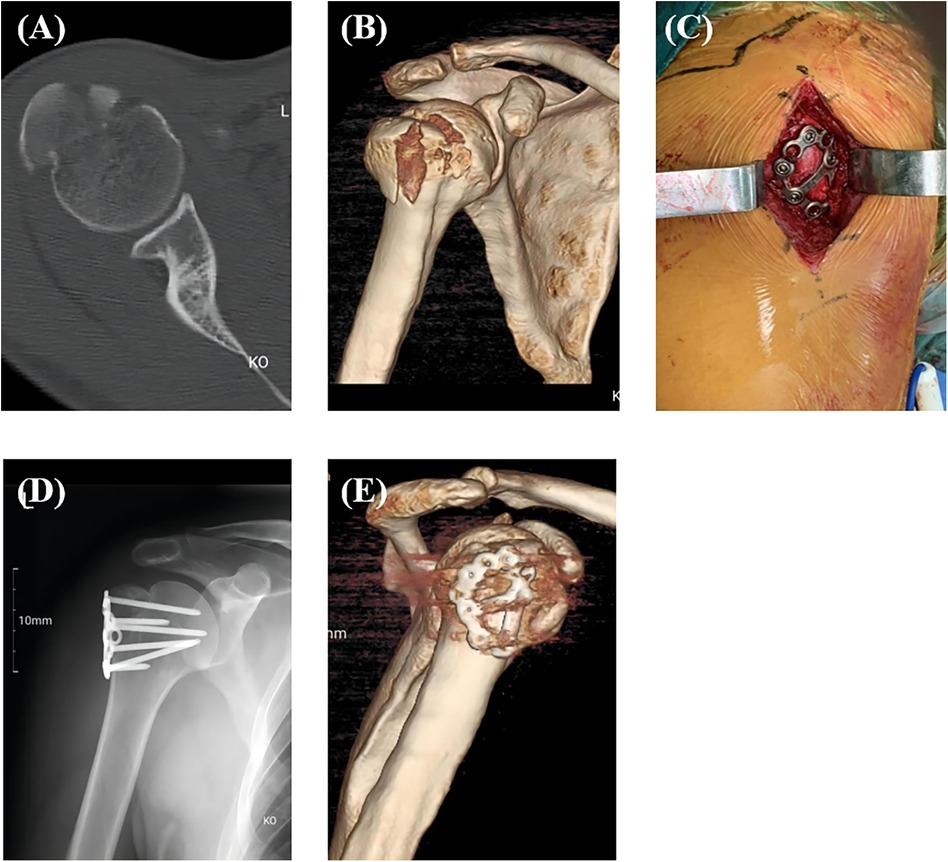
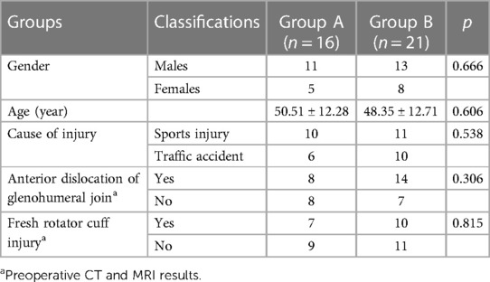
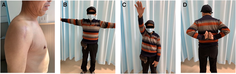
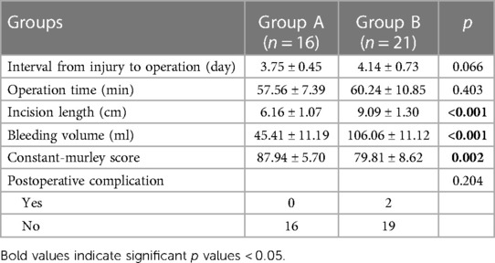
Novel calcaneal plate versus traditional philos plate for treating split fractures of humeral greate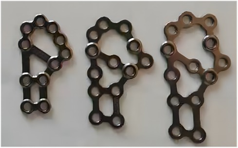 Department of Orthopaedics, Lishui City People’s Hospital, The Sixth Affiliated Hospital of Wenzhou Medical University, The First Affiliated Hospital of Lishui University, Lishui, Zhejiang, China Purpose: Patients and methods: Results: Conclusion: Fractures of the humeral greater tuberosity occur typically in young patients with high-energy trauma, especially in males (1–3), accounting for about 20% of proximal humeral fractures (4, We hypothesized that a calcaneal anatomical locking plate can adequately stabilize split fracture of humeral greater tuberosity and prevent the complication of internal fixation failure and impingement syndrome.The aim of this study is to present the calcaneal anatomical locking plate fixation for split fractures of humeral greater tuberosity and evaluate its clinical outcomes. Patients From September 2016 to April 2021, a total of 37 patients with split fractures of humeral greater tuberosity were treated in our hospital. Patients with fracture and dislocation underwent a routine manual reduction in emergency, and were given symptomatic treatment such as limb immobilization, detumescence and analgesia after admission. The interval from injury to operation ranged from 1 to 7 days. All operations were performed by the same surgical team. This retrospective study was approved by the institutional review board of Lishui City People's Hospital. All participants' written informed consent were obtained before its commencement. We reported this study in accordance with the Helsinki Declaration and following the Strengthening the Reporting of Cohort Studies in Surgery (STROCSS). Inclusion criteria were as follows: (1) patients aged >18 years old; (2) patients with split fracture of the humeral greater tuberosity according to the classification for greater tuberosity fractures of the proximal humerus proposed by Mutch et al. (8) in 2014; (3) the displacement of the main bone mass of the greater tuberosity >5?mm or the angulation >45° according to preoperative CT; and (4) patients with postoperative follow-up >10 months. Exclusion criteria were as follows: (1) patients with old fracture, or pathological fracture, or open fracture; (2) patients with Bankart injury or Hill-sachs injury as well as ipsilateral nerve and vascular injury; (3)patients with other chronic diseases that affected the function of the ipsilateral shoulder joint; and (4) patients with a previous history of surgery around the ipsilateral shoulder joint. Surgical techniques After successful general anesthesia, patients were adjusted to keep their beach chair position with the shoulder of the affected side padded. In Group A, the longitudinal incision was made from the inferoanterior part of the acromion (deltoid space approach). The standard thoracic deltoid approach was used in Group B. Attention was paid to careful separation during the operation to avoid damaging the axillary nerve and the feeding vessels of the humeral head. The fracture end of the greater tuberosity of the humerus was explored and cleaned to avoid soft tissue embedding, followed by the exposure and exploration of the rotator cuff insertion by rotating the shoulder joint. Rotator cuff suture traction combined with bone tenaculum can be used to reduce the fracture end, and then one or two 1.5?mm Kirschner wires were used to temporarily fix the fracture end. After the identification of the good position of the fracture end under C-arm fluoroscopy, the calcaneal plate was trimmed to cut off the excess part according to the size of the fracture fragments in Group A, followed by plastic processing and then implantation. Different sizes of anatomical plates are available (Figure?1). In Group B, the PHILOS plate was inserted via the interpectoralis major and deltoid groove approaches. The implantation position of the plate was that the upper edge should not exceed 3?mm below the apex of the greater tuberosity, and the leading edge should be about 5?mm outside the intertubercular sulcus. Screwing was performed according to the shape and position of the fracture. According to the rotator cuff injury, the injury was repaired with a non-absorbable suture, which can be sutured on the edge of the plate or screw hole through the bone-plate gap. Finally, the fracture reduction, internal fixation position and length were confirmed to be satisfactory under x-ray. The shoulder joint was mobilized to avoid subacromial impingement, followed by routine placement of the drainage rubber and suture of the incision layer by layer. A typical case in Group A was shown in Figure 1 FIGURE 1. Calcaneal plates of different sizes. Figure 2 FIGURE 2. A typical case in group A. Preoperative CT (A) and 3D reconstruction (B) of the split fracture of humeral greater tuberosity; Macroscopic picture after placement of the calcaneal anatomical plate (C); Postoperative x-ray (D) and 3D reconstruction (E). Postoperative rehabilitation After the operation, patients were informed to use the forearm sling for 4–6 weeks initially, with elbow, wrist, and finger activities within the allowable range. The shoulder joint can swing and restrict to internal rotation to protect the fracture fragment. Patients were permitted to have passive activities 1–2 weeks after operation. According to the results of imaging examination at 4 weeks after operation, the fracture healing was judged, and active assistance and active mobilization were gradually started. Weight-bearing activities could be performed 10–12 weeks after operation. In addition, patients could choose to take out the implant 12 months after operation. Assessments Basic clinical information, injury-related variables, interval from injury to operation, incision length, operation time, bleeding volume and postoperative Constant-Murley shoulder joint function score, and postoperative complication were recorded. The Constant-Murley shoulder joint function score was used for evaluation of shoulder joint activity function. including pain scale 15 points, muscle strength 25 points, joint range of motion 40 points, and daily life 20 points (9). The higher the score, the better the shoulder joint function. Postoperative complications mainly include axillary nerve injury, fracture reduction loss, bone nonunion, postoperative infection, ischemic osteonecrosis, shoulder impingement syndrome, and others. Statistical analysis The SPSS 22 software was used for all statistical analyses. was used to compare the variables in two groups. Quantitative variables were shown in the form of mean?±?standard deviation and independent sample Sixteen patients were treated with calcaneal anatomic locking plate (Group A) and 21 with Proximal Humeral Internal Locking System (PHILOS) (Group B) for internal fixation. There were 24 males and 13 females, with an average age of 46.7 years old (ranging from 25 to 67). In terms of the causes of injury, there were 21 cases of falls and sports injuries, and 16 cases of traffic accident injuries. According to the preoperative CT and MRI results, 22 cases were accompanied by anterior dislocation of the glenohumeral joint, and 17 cases had fresh rotator cuff injury (Table?1). Table 1 TABLE 1. Comparison of demographics and injury-related variables between two groups. All 37 cases had fracture healing during the follow-up period (ranging from 10 to 23 months), with an average of 11.9 months. No significant difference was observed in the average interval from injury to operation between Group A and Group B. The average length of incision in Group A was significantly shorter than that of Group B. There was no significant difference between the average operation time of the two groups (p?=?0.403). There was a significant difference in the comparison of bleeding loss between Group A and Group B (Group A, 45.41?±?11.19?ml; Group B, 106.06?±?11.12?ml, Figure 3 FIGURE 3. One case in group A with good shoulder function at 10 months after operation. Small surgical incision (A); Shoulder abduction (B); Shoulder flexion (C); Shoulder external rotation (D). Table 2 TABLE 2. Comparison of surgery-related variables, constant-murley score and postoperative complication between two groups. Approximately 90% of the fractures of the greater tuberosity of the humerus had no or slight displacement and can be managed conservatively (10). However, split fractures of humeral greater tuberosity have different characteristics to other proximal humeral fractures. Thus, treatment for split fractures of humeral greater tuberosity should be different to that of other proximal humeral fractures (11). Although indications for surgical treatment of split fractures of humeral greater tuberosity remain controversial, the most common surgical criterion is the displacement of the main bone fragment >5?mm and/or the angulation >45° (3, At present, there are multiple therapeutic choices for the surgical treatment of split fractures of humeral greater tuberosity. Arthroscopic and open approach treatments have been proposed and applied clinically, including transosseous suture fixation, tension band wires, anchors, simple screws, various plate fixation, and arthroscopic double-row suture anchors. However, it is still disputed with regard to the optimal treatment of displaced fractures (13, At present, PHILOS has been widely used in the surgical treatment of split fractures of humeral greater tuberosity (12). Because the PHILOS plate is thick, rigid and difficult to plastic, it is not a good match for the fixation of split fractures of humeral greater tuberosity. In addition, the traditional anatomical locking plate of the proximal humerus is designed with the proximal directional screw inserted into the humeral head. The proposed directional and non-adjustable insertion trajectory cannot achieve adequate fixation for various split fracture types of humeral greater tuberosity, especially for comminuted fracture types. Occasionally, implantation of this anatomical plate in a more proximal position to capture more fracture fragments may increase the risk of shoulder impingement (19). As proposed by many scholars, various mini-locking plates could be used to treat fractures of the greater tuberosity (14, By reviewing the advantages and disadvantages of various implants and the local anatomical characteristics of the humeral greater tuberosity, we found that calcaneal plate is one of the best options for the treatment of split fractures of humeral greater tuberosity. To be specific, compared with the traditional proximal humeral locking plate, the calcaneal mesh anatomical locking plate has a lower profile, better elasticity and thinner thickness, which can facilitate intraoperative plastic processing and trimming. Considering the anatomical morphology of the humeral greater tuberosity, the contralateral calcaneal plate may have better compatibility and stability after pre-springing and plastic processing. Different sizes of plates are available to cover and fix fractures at any part of the humeral greater tuberosity, especially for comminuted fractures. Owing to the small size of the calcaneal plate, it is feasible to adopt the simple subacromial deltoid space approach. The size of the calcaneal plate will be smaller after trimming based on the size of the fracture fragment, leading to no requirement for intraoperative exposure of the axillary nerve. Compared with the traditional proximal humeral locking plate, the calcaneal mesh plate can be used jointly with a variety of screws to fix the humeral head and humeral axis in multiple planes, capture separate fragments, and increase the stability of the structure. When the calcaneal mesh plate is fixed on the humeral greater tuberosity, it can contain the soft tissue and rotator cuff, just like a string bag, which can increase the stability of the structure in a way that only screws cannot achieve. For some patients with rotator cuff injury, it allows the suture for repairing the rotator cuff to be bound to the plate to strengthen the fixation, without impact on the blood supply of the rotator cuff (23, The present study showed that the calcaneal anatomical locking plate has the advantages of less surgical trauma, less bleeding, and better shoulder joint function. Additionally, this type of plate can reduce the incidence of shoulder impingement syndrome. Overall, the surgical technique of using calcaneal anatomical locking plate for the treatment of split fractures of humeral greater tuberosity is easy and efficient. The present study has some limitations. First, this study was performed based on a small sample size. Second, due to the short follow-up time, the implants were not completely removed in all patients at the last follow-up, which may produce a negative impact on the shoulder joint function. Third, biomechanical tests and finite element analysis of calcaneal plates for the treatment of split fractures of the humeral greater tuberosity will be performed in the future. Calcaneal anatomical locking plate may provide a new choice for the surgical treatment of split fractures of humeral greater tuberosity as it has the advantages of less surgical trauma, less bleeding, and better shoulder joint function. The raw data supporting the conclusions of this article will be made available by the authors, without undue reservation. The studies involving humans were approved by Ethics committee of Lishui City People's Hospital. The studies were conducted in accordance with the local legislation and institutional requirements. The participants provided their written informed consent to participate in this study. Written informed consent was obtained from the individual(s) for the publication of any potentially identifiable images or data included in this article. FW: Conceptualization, Data curation, Formal analysis, Investigation, Methodology, Project administration, Validation, Writing – original draft. XN: Data curation, Formal analysis, Investigation, Methodology, Writing – review & editing. HX: Data curation, Investigation, Methodology, Writing – review & editing. WL: Formal analysis, Investigation, Methodology, Writing – review & editing. ZH: Investigation, Methodology, Writing – review & editing. JL: Conceptualization, Investigation, Project administration, Supervision, Writing – review & editing. The author(s) declare that no financial support was received for the research, authorship, and/or publication of this article. The authors declare that the research was conducted in the absence of any commercial or financial relationships that could be construed as a potential conflict of interest. All claims expressed in this article are solely those of the authors and do not necessarily represent those of their affiliated organizations, or those of the publisher, the editors and the reviewers. Any product that may be evaluated in this article, or claim that may be made by its manufacturer, is not guaranteed or endorsed by the publisher. 1. Kristiansen B, Barfod G, Bredesen J, Erin-Madsen J, Grum B, Horsnaes MW, et al. Epidemiology of proximal humeral fractures. PubMed Abstract 2. Omid R, Trasolini NA, Stone MA, Namdari S. Principles of locking plate fixation of proximal humerus fractures. PubMed Abstract 3. Gruson KI, Ruchelsman DE, Tejwani NC. Isolated tuberosity fractures of the proximal humeral: current concepts. PubMed Abstract 4. Bahrs C, Lingenfelter E, Fischer F, Walters EM, Schnabel M. Mechanism of injury and morphology of the greater tuberosity fracture. PubMed Abstract 5. Green A, Izzi J Jr. Isolated fractures of the greater tuberosity of the proximal humerus. PubMed Abstract 6. Liao W, Zhang H, Li Z, Li J. Is arthroscopic technique superior to open reduction internal fixation in the treatment of isolated displaced greater tuberosity fractures? PubMed Abstract 7. Mattyasovszky SG, Burkhart KJ, Ahlers C, Proschek D, Dietz SO, Becker I, et al. Isolated fractures of the greater tuberosity of the proximal humerus: a long-term retrospective study of 30 patients. PubMed Abstract 8. Mutch J, Laflamme GY, Hagemeister N, Cikes A, Rouleau DM. A new morphological classification for greater tuberosity fractures of the proximal humerus: validation and clinical implications. PubMed Abstract 9. Constant CR, Murley AH. A clinical method of functional assessment of the shoulder. Crossref Full Text 10. Platzer P, Kutscha-Lissberg F, Lehr S, Vecsei V, Gaebler C. The influence of displacement on shoulder function in patients with minimally displaced fractures of the greater tuberosity. PubMed Abstract 11. Kim E, Shin HK, Kim CH. Characteristics of an isolated greater tuberosity fracture of the humerus. PubMed Abstract 12. Ortmaier R, Filzmaier V, Hitzl W, Bogner R, Neubauer T, Resch H, et al. Comparison between minimally invasive, percutaneous osteosynthesis and locking plate osteosynthesis in 3-and 4-part proximal humerus fractures. PubMed Abstract 13. Rouleau DM, Mutch J, Laflamme GY. Surgical treatment of displaced greater tuberosity fractures of the humerus. PubMed Abstract 14. Bogdan Y, Gausden EB, Zbeda R, Helfet DL, Lorich DG, Wellman DS. An alternative technique for greater tuberosity fractures: use of the mesh plate. PubMed Abstract 15. Dimakopoulos P, Panagopoulos A, Kasimatis G. Transosseous suture fixation of proximal humeral fractures. PubMed Abstract 16. Niall DM, O’Mahony J, McElwain JP. Plating of humeral shaft fractures–has the pendulum swung back? PubMed Abstract 17. Scheibel M, Lichtenberg S, Habermeyer P. Reversed arthroscopic subacromial decompression for massive rotator cuff tears. PubMed Abstract 18. Gaudelli C, Ménard J, Mutch J, Laflamme GY, Petit Y, Rouleau DM. Locking plate fixation provides superior fixation of humerus split type greater tuberosity fractures than tension bands and double row suture bridges. Crossref Full Text 19. Südkamp N, Bayer J, Hepp P, Voigt C, Oestern H, K??b M, et al. Open reduction and internal fixation of proximal humeral fractures with use of the locking proximal humerus plate. Results of a prospective, multicenter, observational study. Crossref Full Text 20. Xue G, Chahal K, Lim T, Hu S, Li S, Liu J. Titanium mini locking plate with trans-osseous sutures for the treatment of humeral greater tuberosity fracture osteosynthesis versus PHILOS: a retrospective view. PubMed Abstract 21. Clavert P, Adam P, Bevort A, Bonnomet F, Kempf JF. Pitfalls and complications with locking plate for proximal humerus fracture. PubMed Abstract 22. Theopold J, Marqua? B, Fakler J, Steinke H, Josten C, Hepp P. The bicipital groove as a landmark for reconstruction of complex proximal humeral fractures with hybrid double plate osteosynthesis. PubMed Abstract 23. Sch?ffl V, Popp D, Strecker W. A simple and effective implant for displaced fractures of the greater tuberosity: the “bamberg” plate. Crossref Full Text 24. Szyszkowitz R, Seggl W, Schleifer P, Cundy PJ. Proximal humeral fractures. Management techniques and expected results. Crossref Full Text Keywords: Citation: Received: Published: Edited by: Xun Sun, Tianjin Hospital, China Reviewed by: Feng Rao, Peking University People's Hospital, China Zhenjiang Ma, Shanghai Jiao Tong University, China ? 2024 Wang, Niu, Xia, Liang, Hu and Lan. This is an open-access article distributed under the terms of the *Correspondence: Disclaimer:
|

Shanghai Carefix Medical Instrument Co., Ltd.China
滬ICP備16042301號-1 (滬)-非經(jīng)營性-2016-0122 滬食藥監(jiān)械生產(chǎn)許20101725號
