
An external fixator is a surgical device that allows a fractured or misaligned bone to heal properly. By immobilizing the bones, the external fixator provides stability to bone and soft tissue. The device is commonly used to help children heal or to protect skin that is damaged around the bone.
This article explains when and how an external fixator is used, the types of devices, and how to avoid infection or other possible risks.
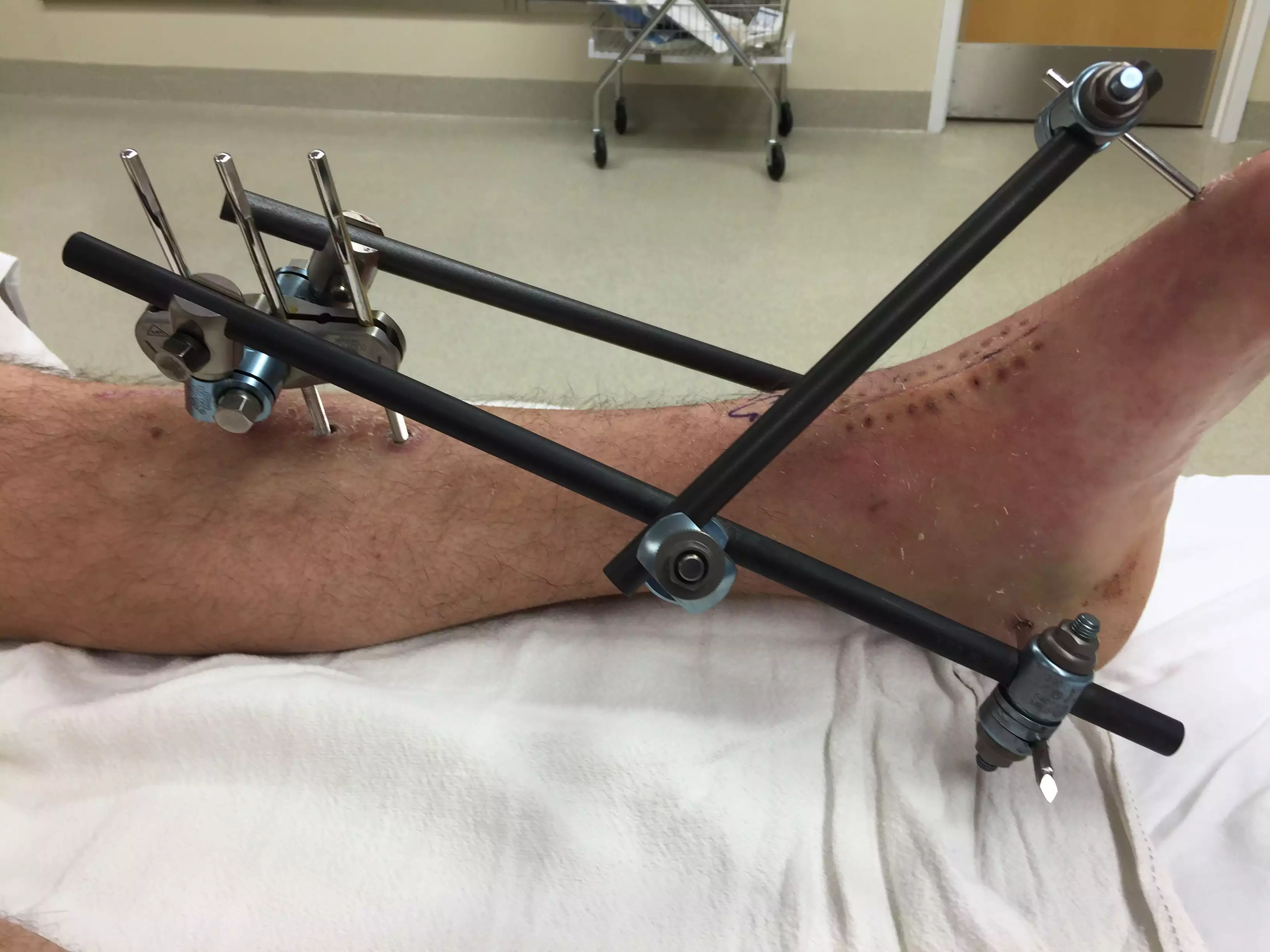
External Fixation to Repair Broken Bone
External fixation is accomplished by placing pins or screws into the bone at various points. In fractures, the pins are typically placed above and below the fracture. The pins are secured together outside of the skin using a series of clamps and rods known as the external frame.
Benefits of an External Fixator
The main advantage of external fixation is that it is quickly and easily applied. The risk of infection at the site of the fracture is minimal, although there is a chance of infection where the rods go through the skin.
External fixators are often used in severe traumatic injuries. They allow for rapid stabilization while allowing access to soft tissues that may also need treatment. This is particularly important when there is significant damage to skin, muscle, nerves, or blood vessels.
Fractures
External fixation ensures the ideal compression, extension, or neutralization of bone placement while allowing for movement of the nearby joints. This aids in setting the bones correctly and can also help minimize muscle atrophy and edema (the buildup of excess fluid) caused by the total immobilization of a limb.1
Limb Alignment and Lengthening
External fixators can help with limb lengthening if one leg is shorter than the other and alignment if a child has a condition such as bowed legs. In these procedures, the external fixator is placed and a surgical procedure is done to make space for new bone to grow.
After the procedure, you will need to turn the struts daily to help lengthen the bone. Your child will also have stretching and strengthening exercises they will need to complete daily. Your care team will instruct you how often your child needs to come back for follow-up, help set up physical therapy appointments, and provide any additional instructions, such as pin care to avoid infection.2
Other Uses
External fixators are also used in other procedures. Two examples are arthrodesis procedures and the treatment plan for osteomyelitis. Arthrodesis is a joint fusion procedure. External fixation can help stabilize the joint, allowing it to fuse.3
Osteomyelitis is a bone infection that can occur due to trauma. External fixation helps stabilize the bone so that it can heal.4
External fixation can also be used to retain the integrity of bone structures (such as the hand) after a serious burn or injury. Without fixation, the exposed or damaged tissue can contract from the accumulation of scar tissue, causing long-term or even permanent restriction of movement.5
Contraindications for External Fixation
External fixation is contraindicated under the following circumstances:6
Bone-related disorders or deterioration that make stabilization less assured.
Persons who are not able or willing to properly care for the pins and wires.
A person with a severely compromised immune system who are at higher risk of infection.
How an External Fixator Is Applied
Orthopedic surgeons do external fixation procedures under general anesthesia. The procedure itself typically follows the following steps:
Holes are drilled into the undamaged areas of bones around the fracture, infection, or area for joint infusion.
Special bolts are screwed into the holes.
Outside of the body, rods with ball-and-socket joints are joined with the bolts.
Adjustments are made to the ball-and-socket joint to ensure the bone is aligned properly with as little, if any, shortening of a bone.
The bolts and external frame can usually be removed without anesthesia in a healthcare provider's office. Fractures have been known to occur at the drill sites, so extended protection may be needed after the device is removed.
Types of External Fixators
There are several different types of external fixator devices. The two main types are circular external fixators and monolateral external fixators.7
Circular external fixators: Circular external fixators go around the limb being treated. They have two rings at the top and bottom connected by struts, wires, or pins. They look cylindrical.
There are two main types of circular external fixators. They are the Taylor spatial frame (TSF) and the Ilizarov external fixator. The TSF is more commonly used than the Ilizarov. Both require adjustments to achieve the desired length of the bone.
The Ilizarove fixator requires a wrench to adjust the device, and the TSF is more easily adjusted. However, the Ilizarove is sometimes the preferred design for small children.7
Monolateral external fixators: A monolateral external fixator is a straight bar that runs parallel to the treated bone instead of going around the bone. Screws are placed in the bone. The screws are often coated in hydroxyapatite so that they adhere to the bone better. These fixators have knobs on them that may need adjusting if the goal is lengthening the bone.7
Risk of Infection
The main risk of infection is at the pin sites where the external fixator is connected to the bone since this is where broken skin is present. To help prevent infection, following the pin care instructions from your healthcare team is essential. They will explain how to care for the pin sites and tell you how often you should clean them — typically once per day.
Your healthcare team will provide you with specific instructions. However, pin care generally includes the following steps:8
Wash your hands and put on clean gloves.
If there is any dry discharge around the pins, wet pieces of gauze and place a separate piece around each pin site.
Use a separate cotton swab at each pin site to remove any dried discharge once it has softened under the gauze.
Use new, separate cotton swabs to clean each pin site if there is any additional drainage.
Clean each pin site with a bottle of sterile saline.
Use gauze or cotton swabs to dry each pin site. Use a new piece of gauze or a new cotton swab for each pin site.
It is important to note that germs from one pin site can contaminate another. So, to help prevent infection, make sure to use a new piece of gauze or cotton swab for each pin site you are working with.
Signs of Infection
Some redness and crusting around the pin sites are expected. However, if you notice the pin sites are redder or swollen, they feel more painful, there is pus, or you develop a fever, contact your healthcare team for advice. You may need antibiotics to clear an infection.
Summary
External fixators allow a healthcare provider to stabilize a broken bone quickly. They are especially useful in traumas. They are also beneficial in correcting limb alignment or when a limb needs to be lengthened. External fixators can serve other uses, too, such as in joint fusions. Several different types of external fixators exist, and your healthcare provider will use the one best suited to your condition.
While the risk of infection is minimal, a pin site infection is possible. Make sure to follow the instructions from your healthcare team and contact your provider if you are concerned you may have developed an infection.


